
The Imaging platform of the Radiology Department at Munich University Hospital
Imaging techniques play an important role in diagnosing pathological lung changes. Whether microscopy, computed tomography or magnetic resonance imaging – the images facilitate and improve diagnosis and enable more precise treatment as early as possible.
Advances in computed tomography (CT), the reduction of radiation dose and the improvement of magnetic resonance imaging (MRI) have already led to a significant improvement and increased depth of imaging.
The DZL promotes research in the field of thoracic radiology. The aim is to open up new fields of indication for other disease areas. Research is also being carried out into how doctors can use imaging to differentiate more precisely between malignant and benign lung diseases. The Radiology Imaging Platform at the Munich University Hospital provides state-of-the-art techniques and equipment for this purpose and thus supports research in the DZL and beyond.
Current projects in the field of radiological imaging:
Precise detection of Covid-19 lung defects using low dose CT
Lead by Prof. Dinkel a team of researchers around Dr. Mircea Stoleriu explored the possibility of using low dose Computed Tomography in conjunction with a dedicated texture analysis (IMBIO CT Lung Tecture AnalysisTM) to measure the severity of COVID-19-induced lung pathologies and prediction of disease course along with clinical parameters:
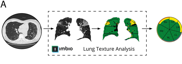
© LMU Klinikum
Non-invasive functional lung imaging
Fourier Decomposition MRI (FD-MRI) offers a unique oportunity to measure and depict region functional lung parameters (perfusion and ventilation) without using any external contrast material whatsoever. However, patient-related uncertainties such as irregular breathing and/or an irregular heart beat decrease both quality and accuracy of the desired parameters. To overcome these obsticles the team of Prof. Dinkel has teamed up with the University Hospital Basel. By employing modern signal processing tools first results have shown the possibility to compensate for these irregularties:
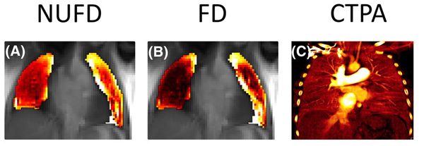
© LMU Klinikum
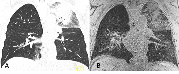
MRI to replace CT in immunosuppressed patients
Pneumonia in immunosuppressed patients is a common complication that can be fatal despite modern prophylaxis. While computer tomography offers good sensitivity in the detection pneumonia, many immunosuppressed patients are young and affected by hematological disease with a very good prognosis. Therefore, the cumulative CT induced radiation dose is a great concern in these patients. Using modern MRI techniques, in collaboration with Siemens Healthcare researcher Shiwa Mansournia explores the feasibility of MRI in these patients:
Pulmonary imaging in radiation Oncology
Radiologic imaging plays a major role in radiation therapy to delineate tumour boundaries and to monitor disease after treatment. Due to its excellent soft tissue contrast and absence of ionizing radiation, MRI is an ideal candidate to improve radiation therapy by image guidance. New hybrid machines, such as the MR-Linac, aim to make use of this synergy by combining an MRI-system with a medical linear accelerator. However, image guidance of radiation therapy in the chest is further complicated due to respiratory motion. In initial experiments a team of researchers, comprising both members of the radio-oncology department (Prof. Guillaume Landry) and the radiology department (Prof. Julien Dinkel) have investigated the MRI-based uncertainties in planning the treatment of lung tumours:
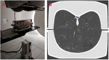
© LMU Klinikum
![[Translate to Englisch:] Frühgeborenes mit Lungenkrankheit](/fileadmin/CPC-M/Forschung/Fruehgeborenes-mit-BPD-3.jpg "[Translate to Englisch:] Frühchen mit Lungenkrankheit")